Posterior Tibial Tendon Dysfunction w/ Flat Foot Reconstruction Options

What is Posterior Tibial Tendon?
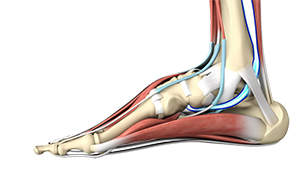
The posterior tibial tendon is responsible for facilitating for patient’s to get up on their toes. It runs from the back of the calf around the inside of the ankle to the arch of the foot. It provides stability to the arch and supports the foot while walking. Inflammation or a tear of this tendon as a result of injury may cause dysfunction, leading to pain and the development of a flatfoot. Posterior tibial tendon (PTT) syndrome or insufficiency is common in all age groups but often middle age patients with flat feet are most symptomatic.
Symptoms
Symptoms of flatfoot include pain on the inside of the ankle that may be accompanied by swelling. Flatfoot may cause the heel to shift outwards causing pain on the outside of the ankle as well. Activities such as walking, running, and standing on your toes may aggravate pain. If treatment is delayed there may be rigidity and development of arthritis. You may have trouble walking or wearing shoes.
Non- Surgical Treatment
Posterior tibial tendon dysfunction in its early stages may be treated with
- Orthotics: Orthotics are designed to help alleviate the mechanical malalignment and are often the first line of treatment.
- Our department has an on-site fully trained orthoptist to help design the best orthotic for each individual patient.
- Physical Therapy
Surgical Treatments:
Surgery has traditionally been involved with tendon transfers and bone osteotomies that used to take up to one full year to recover. We have pioneered minimally invasive procedures that allow the foot to be realigned with small incisions and allow the tendon regenerate when mild to moderate degeneration.
Surgical Procedures Include:
- Tenosynovectomy: The inflamed tendon tissue is cleaned and removed.
- Tendon transfer: The damaged tendon is replaced by another foot tendon.
- Arthrodesis: In cases where arthritis has developed, the bones are realigned and fused to form a single bone by removing cartilage.
- Osteotomy: Bones of the heel and midfoot may be cut to recreate the arch of the foot.
In Office:
Nano Tendoscopy
This includes:
- Visualizing the PTT through keyhole surgery, debriding the tendon.
- Seeding of stem cells from patient’s iliac crest.
When it is needed:
- Early PTT
In Operating-Room:
1. Nano Tendoscopy (including Bioresis Screw)
This includes:
- Visualizing the PTT through keyhole surgery, debriding the tendon.
- Using the placement of a biorhesis screw through the calcaneus to lift the foot, creating an arch.
- Seeding of stem cells from patient’s iliac crest.
When it is needed:
- PTT
2. All American Procedure
This includes:
- A heel bone slide, tarsal metatarsal fusion, lateral column lengthening as well as tendon transfer and ligament reconstruction.
- Seeding of stem cells from patient’s iliac crest.
When it is needed:
- Grade II or early Grade III PTT
You can get around this if:
- You can unilaterally toe raise and have limited degeneration or tearing of the PTT.
Old Approaches vs. New Approaches
Surgery has traditionally been involved with tendon transfers and bone osteotomies that used to take up to one full year to recover. This so called All-American procedure has excellent outcomes but it is not always required for lower grade PTT deficiencies. Dr. Kennedy has pioneered minimally invasive approach to flatfoot surgery that is less invasive and accelerates recovery to 2-3 months post op. If you are a candidate for the MIS flatfoot reconstruction, the procedure will allow the foot to be realigned with small incisions and allow the tendon to regenerate when mild to moderate degeneration of the tendon is present. Using a nanoscope through a 2mm incision allows us to visualize the PTT and subsequently remove areas of degeneration. Then the patient's own stem cells obtained from their bone marrow can be injected into the tendon to facilitate repair.
Dr. Kennedy's Articles
Functional Outcomes of Tibialis Posterior Tendoscopy With Comparison to Magnetic Resonance Imaging


